Tigger and "Is it IBD or lymphoma?"
Part 2: Coming up with the diagnosis

So to recap, to determine if Tigger had IBD or lymphoma, the steps required were:
After Tigger’s surgery (and steps 1 and 2 completed), I eagerly awaited test results to tell me if I was treating her for IBD or cancer. In theory, it should be that easy, however some diseases (like IBD and lymphoma) can have a blurred line making tissue analysis and final diagnosis a real challenge.
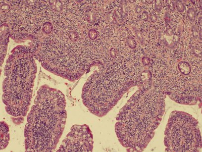
On Tigger’s regular stained sample (a pink/purple stain called H&E), there were lots of lymphocytes (a type of white blood cell appearing as small, dark purple circles) and blunting of the intestinal villi (the finger-like projections that make up the inner lining of the intestine) but not a lot of changes to the structured layers of the intestine itself.
- Get tissue samples
- Send samples to the lab
- Await the pathologist to tell you what disease you are treating
After Tigger’s surgery (and steps 1 and 2 completed), I eagerly awaited test results to tell me if I was treating her for IBD or cancer. In theory, it should be that easy, however some diseases (like IBD and lymphoma) can have a blurred line making tissue analysis and final diagnosis a real challenge.
On Tigger’s regular stained sample (a pink/purple stain called H&E), there were lots of lymphocytes (a type of white blood cell appearing as small, dark purple circles) and blunting of the intestinal villi (the finger-like projections that make up the inner lining of the intestine) but not a lot of changes to the structured layers of the intestine itself.
|
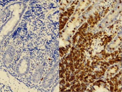
To ensure that all “t”’s were dashed, and all “I”’s dotted (so to speak), I elected to run specially staining on the tissue samples called immunohistochemistry staining. Immunohistochemistry staining is a newer technique where the coloured part of the stain binds only to certain structures. This type of stain allows pathologists to identify between two objects that look identical on regular staining. As most of the inflammatory cells from the first stain were lymphocytes, the special stains were used to identify T lymphocytes versus B lymphocytes. This is where the diagnosis becomes more suggestive for lymphoma. As indicated in the 2 slides, over 95% of the lymphocytes stained positive for T-cell origin.
Why is this important? In inflammatory disease, we expect both T and B cells to be contributing so we should see mixed staining results. As cancers reflect an abnormal growth of one cell type, to see mostly T cells is not good. In Tigger's slides, we can see very few dark blue staining B-cells compared to a sample containing lots of dark brown T-cells. |
To come up with a final diagnosis in this case, one additional molecular technique was run on the tissue sample: polyclonality. Polyclonality is a technique where all the lymphocytes are removed from the processed tissue and the allowed to migrate through a gel block using electric current. The cells can move only as fast as their weight so they will spread out over time. In inflammatory disease the cells are from different sources so they have different weight. An inflammatory gel block will look like a dirty smear. In cancer cells, the cells are coming from the same source so they all weigh the same. This creates distinct bands in the gel.
In Tigger’s case, almost all the cells were of the same weight. This gave us the final diagnosis in her disease: Emerging low grade T-cell lymphoma.
The term emerging lymphoma was used in this case because the structural changes normally seen with classic lymphoma were not observed (the cancer was there, it had just not had enough time to muck around).
Stay tuned to see how Tigger was treated and how she is did with everything…
Click here to go to Part 3: Treatment and Outcome


