Tigger and "is it IBD or lymphoma?"
Part 1: History & Work-up

I have the luxury of using my feline knowledge to watch my cats on a regular basis (my cats may not consider this an added bonus but that is their opinion). This allows me to catch things early but even at times I am caught off guard by the diseases of aging cats.
I adopted Tigger when I first started as a vet assistant in this industry. In my mind, I remember her as the same little kitten I adopted but in reality she has now passed on at the wonderful age of 18 years. I now use her case and the knowledge I gained from caring for her, in guiding how I help other kitties.
In summer 2011, Tigger was eventually diagnosed with low grade alimentary (intestinal) cancer. Given the complicated nature of her disease, I have chosen to break her case up into 3 parts:
- History and work up
- Coming up with the final diagnosis
- Treatment/outcome
History
Tigger was never a “recreational vomiter” – she would have the occasional hairball (i.e. once every 2 – 3 months) and only rarely vomit food so it was quite a shock when she had an episode of vomiting blood in the summer time. Needless to say, vomiting blood warranted an immediate trip to the clinic.
On physical exam, her weight was found to be unchanged and she was not appreciably dehydrated. She was uncomfortable when her abdomen was palpated (examined) and I would have described it as having a very full feeling.
In the abdomen lie intestines, liver, pancreas, stomach, kidneys, and bladder. Some organs (like the kidneys) are easily palpated because they have a defined shape and firm texture. Other organs (like the bladder) can be felt depending on how full they are or (like the liver) should not really be felt (because of location or texture) unless something is wrong.
The intestines are funny structures as they can have content in them (like stool) and be easily felt but small intestines are generally very slippery and not something we can really hold onto unless there is a problem. In Tigger, her intestines were very easily felt, like cords in the belly. This is not a normal finding.
Preliminary Assessment
History and physical exam led me to narrow down my disease concerns to two main differentials:
- Inflammatory bowel disease (IBD)
- Cancer of the intestines, the most common form of which in cats is lymphoma
Imaging to diagnose IBD vs lymphoma
|
X-rays:
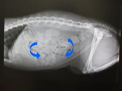
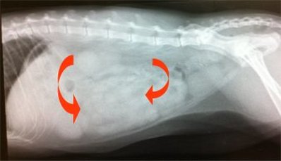
No obvious masses were found on x-ray (big sigh of relief!) but the intestine pattern was not really normal. Normal intestines show mixed pattern of radiodense (grey) areas and radiolucent (black) areas to indicate intestinal content and gas being mixed around during normal digestion. In the normal x-ray (top), the blue arrows indicate gas and digesta filled intestines. Tigger’s xrays (below) show only radiodense areas. The abnormal intestines are marked by red arrows. |
|
Ultrasound:
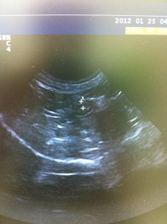
Apart from being able to measure intestinal walls and better visualize soft tissue structures like the spleen, pancreas, and liver, ultrasound shows how structures can move. The challenge with presenting ultrasound images alone is that we miss seeing the movement that comes with this technology. In Tigger’s case, apart from thickened intestines, there was no movement (peristalsis) observed. This condition is known as ileus and is not normal. Thickened intestines occurs in both IBD and cancer so ultrasound proved that something was wrong but that the changes were so far limited to the intestines. |
Plan at this point:
Tigger was started on supportive care to control vomiting, stimulate intestine mobility, and keep her hydrated and pain free while the decision was made to go for surgery or not (she is a family cat so it had to be a family decision).
Surgery was considered as this would provide us with a disease diagnosis. The purpose of surgery was to go in and collect tissue (biopsy) samples of the small intestine and lymph nodes. By analyzing the abnormal intestinal tissue, the goal was to determine if this was a case of IBD or cancer. If the disease did turn out to be cancer, lymph node samples were taken to determine if it had spread (a process called metastasis).
The biggest risks associated with this type of surgery include:
Tigger had surgery about 10 days after her initial vomiting episode and did fantastic. She was kept in hospital for 2 days. The tissue samples were sent off for microscopic analysis (histopathology). This is where things got interesting…
Click here to go to Part 2: Coming up with the diagnosis
Surgery was considered as this would provide us with a disease diagnosis. The purpose of surgery was to go in and collect tissue (biopsy) samples of the small intestine and lymph nodes. By analyzing the abnormal intestinal tissue, the goal was to determine if this was a case of IBD or cancer. If the disease did turn out to be cancer, lymph node samples were taken to determine if it had spread (a process called metastasis).
The biggest risks associated with this type of surgery include:
- Anesthetic recovery: Tigger was deemed a good surgical candidate as she was otherwise in good health. She was maintained on IV fluid support to help keep her blood pressure stable. She was monitored throughout the anesthetic by one of our RVTs.
- Controlling secondary infection: In a case like Tigger’s, the abdomen is clean but cutting into the intestine runs the risk of introducing infection into the abdomen. Good surgical technique along with prudent use of antibiotics reduces this risk to manageable levels.
- Pain management/patient comfort post-op: Surgery is surgery no matter how we look at it. Tigger was maintained on combination narcotic and NSAID pain control for approximately a week post-op. Pain management is important in any case as pain is now considered the 5th vital sign.
Tigger had surgery about 10 days after her initial vomiting episode and did fantastic. She was kept in hospital for 2 days. The tissue samples were sent off for microscopic analysis (histopathology). This is where things got interesting…
Click here to go to Part 2: Coming up with the diagnosis


